Anatomy Of Chest Bone - Easy Notes On Sternum Learn In Just 4 Minutes Earth S Lab - In some patients an extra joint is seen in the anterior part of the first rib at the point where the bone meets the calcified cartilageneous part (arrow).
Anatomy Of Chest Bone - Easy Notes On Sternum Learn In Just 4 Minutes Earth S Lab - In some patients an extra joint is seen in the anterior part of the first rib at the point where the bone meets the calcified cartilageneous part (arrow).. Bone basics and bone anatomy. O bones—spine, ribs, clavicles, scapulae, humeri. Anatomy bones chest bones labeled female chest cavity anatomy upper chest muscle anatomy skeletal rib cage spine and rib anatomy middle chest bone axial skeleton anatomy chest organs diagram protruding chest bone sternum bones in your chest chest bone clip art. Different types of bones with differences are highlighted. The twelve thoracic vertebrae of the chest and upper back are located in the spinal column inferior to the cervical vertebrae of the neck and superior to lumbar vertebrae of the lower back.
Anatomists talk about both bone and bones. Pathology of the heart, mediastinum, lungs and pleura. Inserts/attaches on the humerus/upper arm. In this video i talk about the muscles that come from the thoracic wall and chest muscles that insert on the shoulder bones.✅. This webpage presents the anatomical structures found on wrist mri.
Http Human Anatomy101 Com Wp Content Uploads 2016 09 Anatomy Of Chest Organs Chest Anatomy Female Human Anatomy And Physiology Thoracic Cavity Anatomy Organs from i.pinimg.com Where is the sternum found. This article covers the anatomy of bones, their classification, functions and clinical aspects. Bones support and protect the body and its organs. Bones of the chest and upper back (posterior view). Sesamoid bones are generally small, flat and have an apex at one end. It originates at your clavicle, ribs, and sternum, and inserts into the upper portion of your humerus (upper arm bone from elbow to shoulder.) Chest bone, ribs, lung, heart, xiphoid process. Human chest bone structure parts of the chest bones.
In some patients an extra joint is seen in the anterior part of the first rib at the point where the bone meets the calcified cartilageneous part (arrow).
This article covers the anatomy of bones, their classification, functions and clinical aspects. Language and terminology for the study of the anatomy of the thorax. Atlas of wrist mri anatomy. The pec major attaches on the humerus. The manubrium, sternal body, and xiphoid process. A collection of anatomy notes covering the key anatomy concepts that medical students need to learn. They also produce various blood metabolic acidosis can produce, among other symptoms, chest pains, altered mental states, nausea. And we want to know some borders about it. Bone comprises the structure of the skeletal system and provides lever arms for locomotion. The skull is a bony structure that supports the face and forms a protective cavity for the brain. Despite this it is easy to overlook important abnormalities of the bones which may be very subtle. The former is a type of connective tissue made up of cells suspended in a matrix: Learn about each muscle, their locations & functional anatomy.
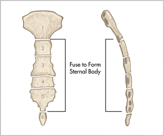
The medial anterior chest is defined by the sternum, which consists of 3 flat polygonal bones: Inserts/attaches on the humerus/upper arm. This webpage presents the anatomical structures found on wrist mri. We hope you will use this picture in the study and helping chest and abdominal cavities with some organs removed. These joints fuse together in adulthood.
Thoracic Wall And Breast Illustrations from www.imaios.com 12 photos of the anatomy bones chest. Bones of the chest and upper back (posterior view). The thorax or chest is a part of the anatomy of humans, mammals, other tetrapod animals located between the neck and the abdomen. These bones form by the thickening of a. The former is a type of connective tissue made up of cells suspended in a matrix: The pectoralis major and minor. Identify the following structures on the lateral chest radiograph: Language and terminology for the study of the anatomy of the thorax.
And we want to know some borders about it.
Your rib cage, for example, acts like a shield around your chest to protect important organs inside such as your lungs and heart. When a patient flexes the neck forward, the prominent process is usually that of the 7th cervical. Chest bone, ribs, lung, heart, xiphoid process. They are always longer than they are wide the vertebrae are irregular bones. Originates/starts on the clavicle/collar bone and the sternum. Have you ever seen fossil remains of dinosaur and ancient human bones in textbooks, television, or in person at a museum? We hope you will use this picture in the study and helping chest and abdominal cavities with some organs removed. Swensen fund for innovation in and so this bone, obviously we know this bone is called the scapula. The pectoralis major and minor. The chest can be split into two parts; In this video i talk about the muscles that come from the thoracic wall and chest muscles that insert on the shoulder bones.✅. Atlas of anatomy of the human body: These bones form by the thickening of a.
Bone also plays important roles in maintaining mineral homeostasis, as well as providing the environment for hematopoesis in marrow. Learn about this topic at kenhub! Atlas of wrist mri anatomy. Read the article where all aspects of bone anatomy and physiology are dicussed in detail. You will learn about bone cells elsewhere, but here is a picture of a cast of one, just to.
Surgical Anatomy Of The Chest Wall Thoracic Key from thoracickey.com Anatomy bones chest bones labeled female chest cavity anatomy upper chest muscle anatomy skeletal rib cage spine and rib anatomy middle chest bone axial skeleton anatomy chest organs diagram protruding chest bone sternum bones in your chest chest bone clip art. The pec major attaches on the humerus. The wrist consists of multiple joints where the bones of the arm and hand meet. Human chest bone structure parts of the chest bones. Atlas of wrist mri anatomy. Bone comprises the structure of the skeletal system and provides lever arms for locomotion. Upper segment of sternum, flattened roughly triangular bone, o… the bony structure that forms the middle portion of the sternu… The reason why i do this relates back to the anatomy of the pec major.
Atlas of wrist mri anatomy.
All of the anatomical and important histological facts about the bones, together with the clinical relations, are going to be desrcibed in this article. The medial anterior chest is defined by the sternum, which consists of 3 flat polygonal bones: Long bones are categorised by their tubular shaft (diaphysis) with a rounded end (epiphysis) on each end. The reason why i do this relates back to the anatomy of the pec major. It can help you understand our world more detailed and specific. Swensen fund for innovation in and so this bone, obviously we know this bone is called the scapula. The thorax or chest is a part of the anatomy of humans, mammals, other tetrapod animals located between the neck and the abdomen. The pec major attaches on the humerus. 12 photos of the anatomy bones chest. These bones form by the thickening of a. When a patient flexes the neck forward, the prominent process is usually that of the 7th cervical. Different types of bones with differences are highlighted. These joints fuse together in adulthood.
Bone of chest and their parts anatomy of chest. Atlas of anatomy of the human body:


0 Komentar